Methods & Technologies
Our lab employs a broad spectrum of methods and technologies for cardiovascular basic research.
From gene to organism.
Our methods cover every level at which cardiac signaling is studied: from sequencing candidate genes to phenotyping transgenic mouse models.
5 method areas · 16 techniques
Genomics & Molecular Biology
Sequencing cardiomyopathy candidate genes, profiling transcriptomes, cloning constructs, and delivering genes with adenoviral or AAV vectors.
- Gene sequencing to identify cardiomyopathy candidate genes
- RNA sequencing (RNA-seq)
- Advanced cloning (Gateway cloning system)
- Adenoviral gene transfer
- AAV-based gene transfer
Proteomics & Interactomics
Profiling the proteome and phosphoproteome of cardiac signaling and detecting protein interactions in situ.
- Proteomics
- Phosphoproteomics
- Proximity ligation assay (PLA)
Cell Culture & Model Systems
Primary cardiomyocytes from neonatal and adult rats, and iPS cells.
- Neonatal rat ventricular cardiomyocytes (NRVCM)
- Adult rat ventricular cardiomyocytes (ARVCM)
- Induced pluripotent stem cells (iPS cells)
Imaging & Microscopy
Localizing proteins and membrane structures in cardiomyocytes.
- Confocal laser scanning microscopy
Immunofluorescence of STRIP1 and nucleolin. Eden / Frey lab. T-tubule network of a cardiomyocyte, ANEPPS membrane stain, 40×. Crop of a single cell, rotated by 90°; original red, shown here in a monotone cyan LUT. Eden / Frey lab.
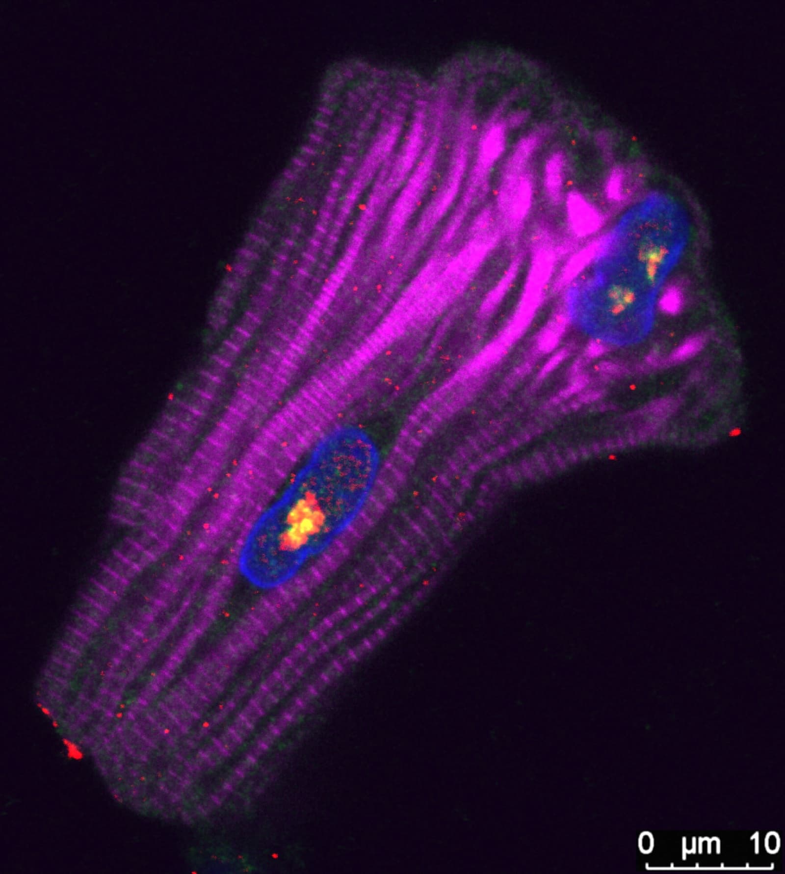 Immunofluorescence
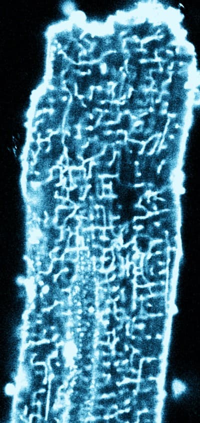 ANEPPS · 40×
In Vivo Phenotyping
Transgenic mouse models, phenotyped by morphometry, echocardiography, and invasive hemodynamics.
- Transgenic mouse models
- Morphometry
- Echocardiography
- Invasive hemodynamics
Research with us.
We are looking for doctoral students, postdocs, and partners who want to apply and advance these methods with us.